Tackling the ‘cowboy field’: transforming glycoproteomics from baby steps to bold strides with Shelley Jager
“Mass spectrometry doesn’t lie. If you don’t get results, it’s because of you. It’s tough love.”
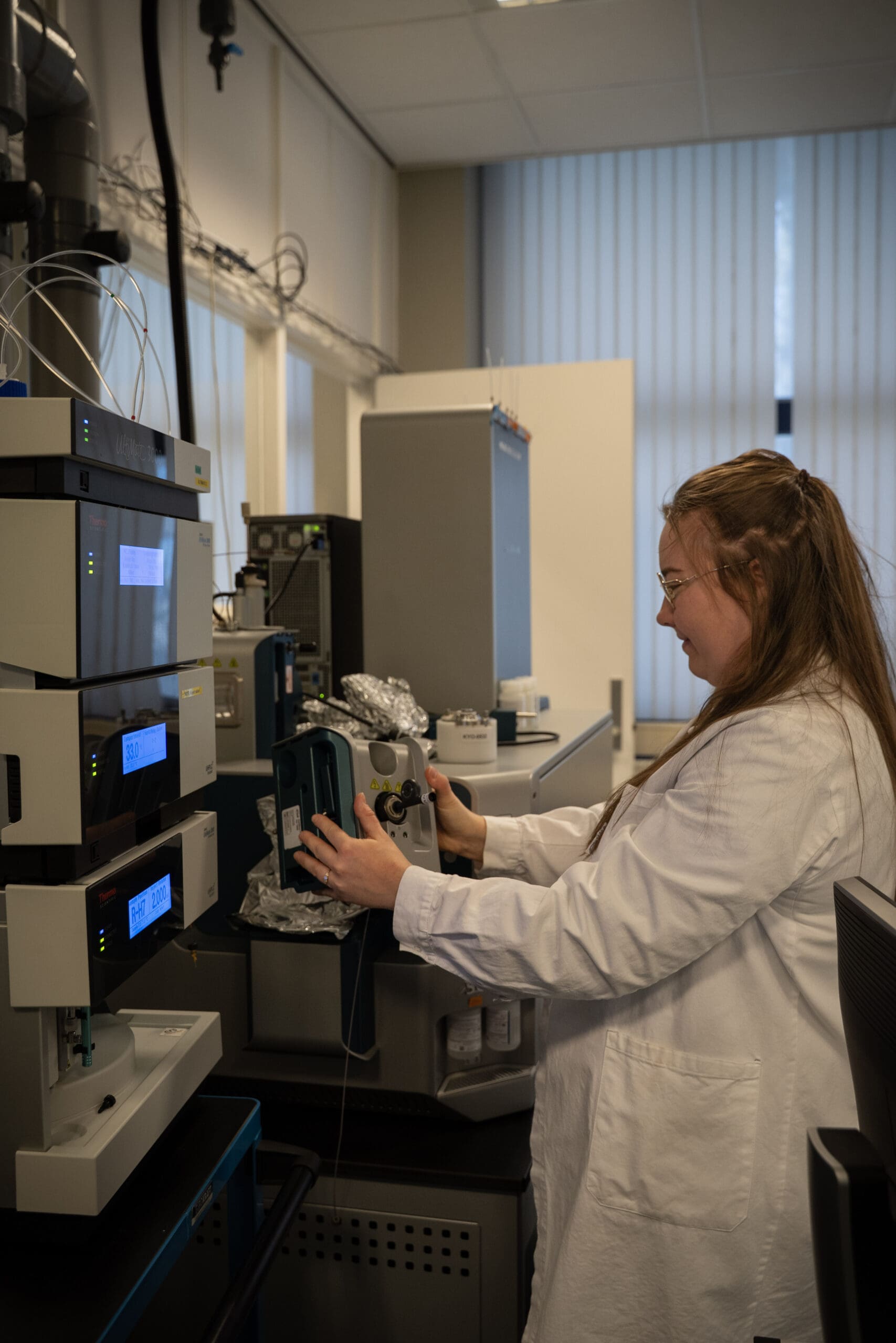
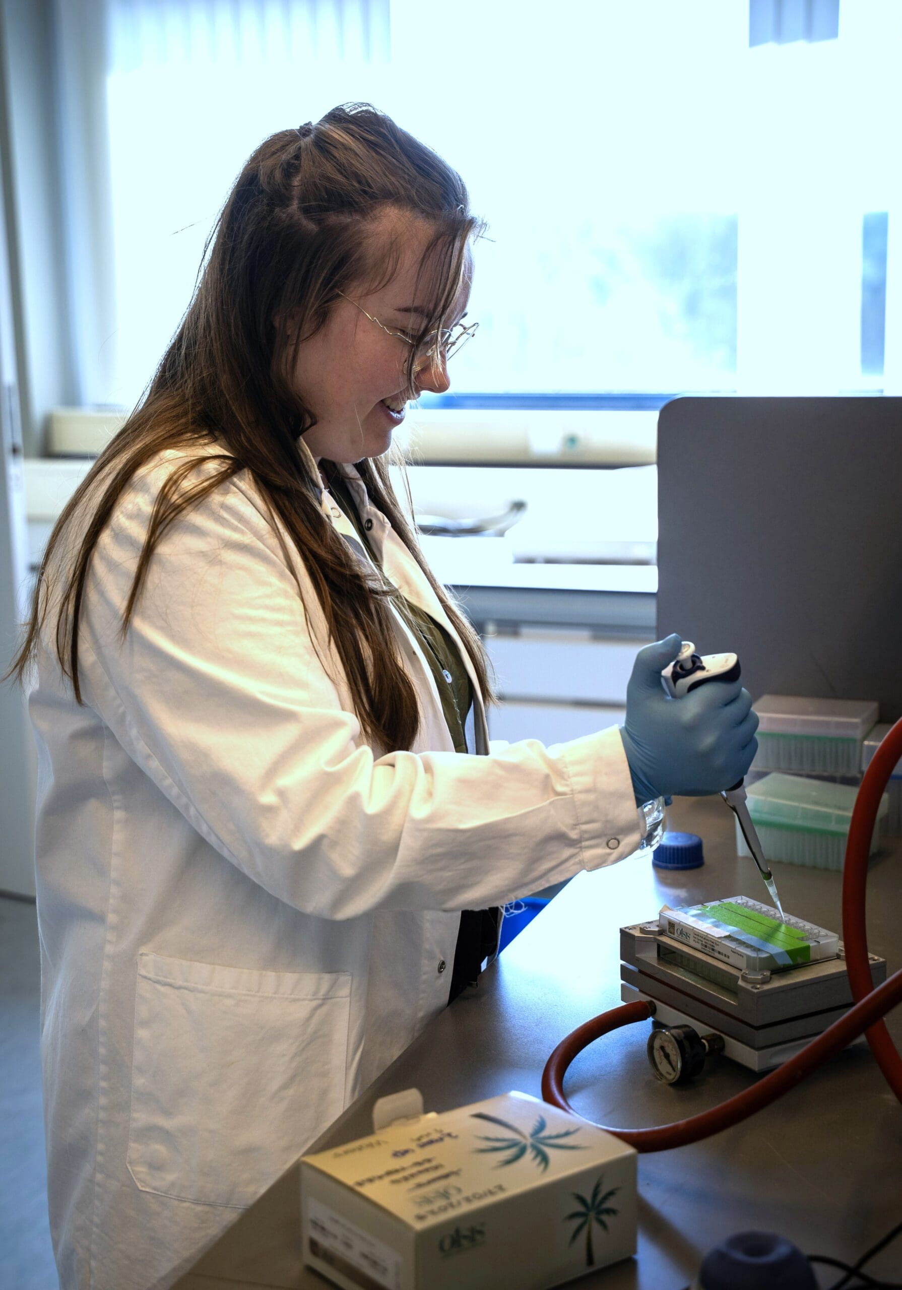
Shelley Jager, PhD candidate in Prof. Albert Heck’s lab at Utrecht University, is pioneering methods in glycoproteomics, a field in its early stages. Initially being an area she wasn’t interested in, Shelley has grown to love the challenge that comes with a largely unexplored field. Every question left unanswered by an experiment is further motivation to keep digging deeper. Her lab’s ‘fun Friday’ tradition provides one unconventional, yet promising pathway to answering such questions.
Tackling the “cowboy field” of glycoproteomics
When Shelley Jager began her PhD at Utrecht University’s Heck lab, she had one request for her supervisor: “Anything except glycoproteomics.” Yet that’s precisely the field she now excels in, having recently co-authored a Nature paper that significantly advances glycoprotein analysis methods.
That initial reluctance transformed into passion as Shelley recognised that glycoproteomics – the study of proteins modified by complex sugar structures – was still in its “baby steps” compared to other proteomics subfields like phosphoproteomics or ubiquitinomics.
“It’s a bit of a cowboy field,” she explains. “I’m finding a challenge in standardising it in my lab, making methods, validating them, getting robust quantification, and pushing the boundaries to get more identifications that we can translate into better biological findings.”
Plasma proteomics: a promising but challenging source
Shelley’s research focuses on developing methods for glycoproteomics, particularly plasma. “For diagnostic purposes, human plasma is a great source, but it comes with many challenges,” she notes. “The dynamic range of proteins in plasma is enormous, which decreases the depth you can reach with mass spectrometry. Glycan heterogeneity further decreases your signal.”
This means that conventional mass spectrometry approaches yield few useful results from plasma samples. “You need really good sample preparation and enrichment,” Shelley explains. “Many existing enrichment methods are quite biased. For example, HILIC enrichment co-purifies many hydrophilic peptides, while ion exchange methods co-purify many non-modified peptides.”
The analytical challenges don’t end there. “It’s not like normal chromatography where you have a nice Gaussian peak,” she says. “Sometimes you see five Gaussian peaks in one peak, at different heights, a little like a heartbeat.” This complexity stems from the three-dimensional nature of glycan modifications, which can branch in different directions and affect the physical properties of the molecule.
From chemistry to fundamental biology
Shelley’s path to glycoproteomics began with an interest in organic chemistry. “Chemistry is nice and fun, but I wanted to go bigger,” she recalls. An internship at the Heck lab, where she is now completing her PhD, introduced her to native mass spectrometry.
“We purified a protein from blood and did native mass spec on it. We found that different people have different proteoform landscapes, and glycosylation was one of the major modifications.” This experience shifted her interest from therapeutic proteins to fundamental biology.
“I always thought it would be nice to develop proteins for therapeutics,” she says. “But that internship showed me there’s already so much to learn about fundamental biology—what changes in a cell to make proteins differently expressed or modified, and what’s the next step in the cascade that changes to result in a different phenotype.”
Overcoming doubt: The Nature paper
Shelley’s recent Nature publication demonstrates how data-independent acquisition techniques can be applied to glycoproteomics, allowing researchers to identify more low-abundance glycoproteins than ever before.
“The reactions were mixed when I proposed data-independent acquisition. Some people said, ‘This is never going to work,'” she remembers. It didn’t help that software was not yet made for data-independent acquisition of glycopeptides. “But one of my supervisors said, ‘Let’s do it.’ And I thought, ‘Oh no, they actually agree—now I have to do it!'”
The project, which took nearly two years from start to finish, yielded remarkable results. “The major highlight is how deep we could go,” Shelley explains. “We found low-abundance cytokines like IL-12, CSF, and other peptides that have never been identified in blood with glycans attached.”
A turning point came when she began using IonOpticks columns for her liquid chromatography separations. “We’d always made our own columns, but when I first used an IonOpticks column on our timsTOF, I noticed something remarkable—glycan isomers were splitting with baseline resolution, not just showing as ugly peaks with a little camel back,” she explains, referencing the improved separation that allowed her to distinguish between structurally similar glycans.
This unexpected benefit transformed her analysis capabilities. “I was amazed at how many isomers we could separate while still getting clean Gaussian peaks instead of these blobs. The peaks are incredibly sharp – even the glyco peaks.”
This discovery of low-abundance cytokines opens new research avenues, as many of these cytokines have typically been studied using proteins produced in E. coli bacteria, which lack the glycosylation machinery found in human cells. “To see that in blood they have glycosylation—not just small glycans, but huge complex glycans that almost double the size of these peptides—was really interesting,” Shelley says.
The implications could be significant, particularly for understanding inflammatory responses. “Cytokines are markers of inflammation. This opens the door to understanding if those glycans also change during inflammation, and if it affects their function.”
Read the full paper here.
The COVID mystery: an unsolved puzzle
Among Shelley’s most intriguing discoveries came during the COVID-19 pandemic, when she analysed blood samples from hospitalised patients. “We noticed these patients had a weird modification on one specific protein that we could only detect with intact measurements,” she recounts. “We saw a mass shift we couldn’t explain, and we only saw it in COVID patients.”
After analysing more than 200 people, including 100 who did not have COVID but had other conditions like cancer, the mysterious modification appeared exclusively in COVID patients. “It was very abundant—about 50% modified, 50% unmodified. Over time, in people who died, the modification increased in abundance, and it decreased in people who survived.”
Despite involving every PhD student and postdoc in the lab and employing every available technique – top-down proteomics, bottom-up proteomics, native mass spectrometry – Shelley and her colleagues couldn’t identify the modification. “It was the most interesting thing I’ve ever found, but we can’t do anything with it because we don’t know what it is,” she says. “I still have these samples in my freezer.”
The team continues to investigate during what they call “Friday Fun Time”—informal experimental sessions where researchers pursue curiosity-driven projects. “Recently we tried to sequence this protein using top-down proteomics on a healthy patient,” Shelley notes. “Interesting findings have come out of Friday Fun Time. I’ve seen people start experiments there that evolved into full projects.”
Making mass spectrometry more approachable
For newcomers to the field, Shelley offers encouragement: “Mass spectrometry is not as difficult as you think. Everyone thinks ‘mass spec is too difficult, I’m not gonna burn my hands on this.’ I would say just do it – it’s intuitive once you understand the basics. That’s what I would say to younger me, who was doubtful that I’d ever understand it.”
Her advice to educators: focus less on hardware and the technique and more on applications. “A lot of lectures are about how ions move through quadrupoles and orbitraps. Even these words are scary for people who don’t know them. Better to start with the applications and the biology that can be achieved with mass spectrometry, and the challenges you can solve that aren’t easily addressed with other techniques.”
For Shelley, the beauty of mass spectrometry lies in its honesty: “It doesn’t lie. If you don’t get results, it’s because of you. It’s tough love.”
The future: isomer separation and glycan structure
Looking ahead, Shelley sees isomer analysis as the next frontier in glycoproteomics. While there are established methods that allow for the study of glycan isomers in glycomics (the study of free glycans), as Shelley explains, “very few people are studying glycan isomers in glycoproteomics [where a peptide is attached to a glycan]. It’s not the same when the antenna are pointing to the right, to the left, away from, or toward each other. These differences have implications in biology.”
Her group now has two researchers focused on translating methods from glycomics to glycopeptides. “The future is looking at these isomers, being able to distinguish them, being able to quantify them separately,” Shelley predicts. “It needs a lot of work still—better software and annotation systems.”
It’s encouraging to Shelley that, with the field getting “hotter”, a lot of this work is already underway. “The isomer separation is key. We can now get separation on C18, without needing a HILIC column that requires a lot of organic solvent and makes the mass spectrometer dirty. Keeping the mass spec cleaner makes me happy, because I’m the one that troubleshoots it if it doesn’t work.”
From a reluctant starter to a passionate advocate pushing the boundaries of a challenging field, Shelley’s journey illustrates how the most rewarding scientific paths can begin with the projects we’re most hesitant to pursue.
Would you like to be featured in an upcoming Community Newsletter? Email us at communications@ionoptickscopy.com and tell us a bit about yourself and your research.